
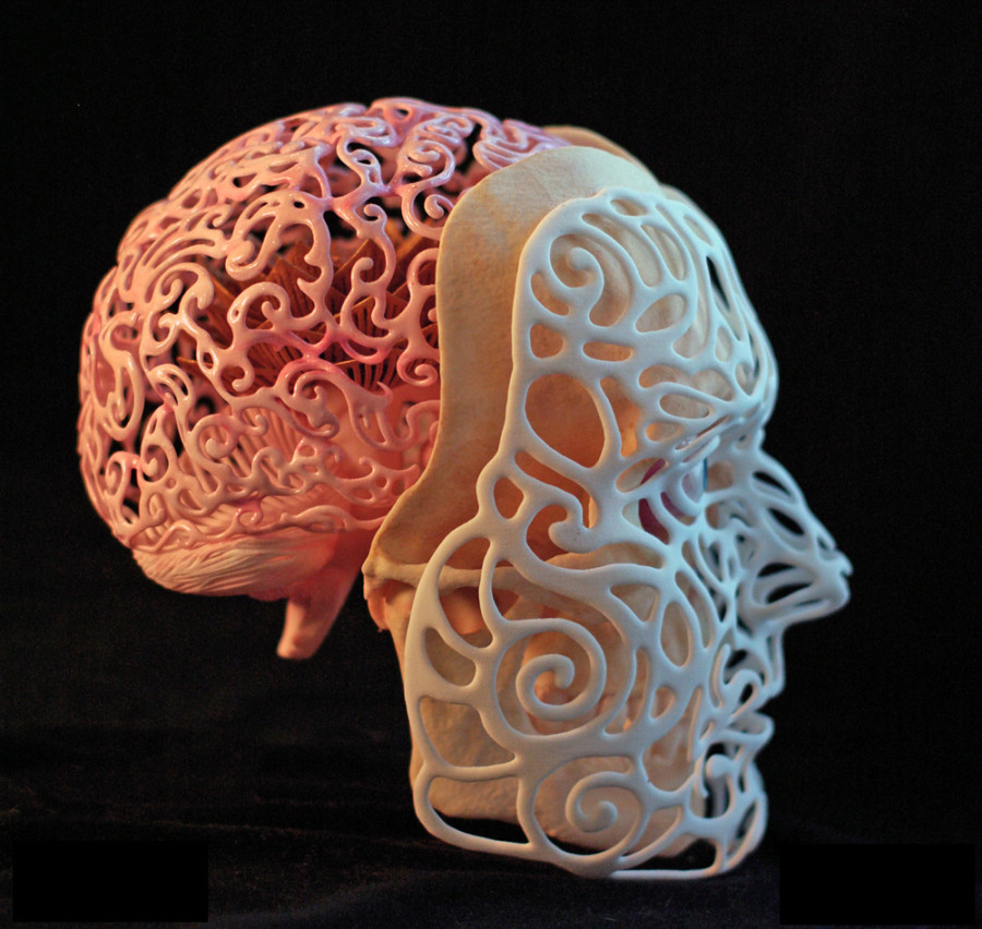

The revolutionary technology of 3D printing has gained traction in the medical field in recent years; spine surgery has in particular seen major advances in 3D printing. The applications of this technology have grown from utilizing 3D models to enhance patient education to patient specific, highly detailed intraoperative anatomical molds.
3D printing and Spine Surgery
Spine surgeons deal with a wide range of complex pathologies of spinal column including deformities, tumours, infections, trauma, and degeneration. All of these problems have different complicated aspects that need to be investigated in detail using various sophisticated techniques. Sound knowledge of anatomy and identification of landmarks, which involve a long and steep learning curve, are some of those crucial requirements for a proper surgical intervention.

Intraoperative techniques for visualization of the deformed spine including preoperative computerized tomography (CT)-based, fluoroscopy-based, and three-dimensional (3D) fluoroscopy have been used as navigating techniques that contribute to the analysis. However, all these techniques involve prolonged surgery duration, high radiation exposure to patients as well as operating staff, and high cost.
Rapid prototyping (RP)/3D printing is a developing approach that sheds light on most of these issues.
How does 3D printing work?
The transfer of the data obtained from 2D imaging to a 3D haptic mannequin constructs this revolution’s basis. This conversion includes miscellaneous available techniques such as stereolithography, selective laser sintering, direct laser metal sintering, two-photon polymerization, laminated object manufacturing, 3D printing, 3D plotting, polyjet inkjet technology, fused deposition modeling, vacuum casting, and milling.
The 3DP’s principle for building up models layer-by-layer with material deposition has led to the impeccable construction of detailed anatomical models or complex, which cannot be reproduced even by sophisticated milling machines. The layering technique includes a liquid that is sprayed through the inkjet printer nozzles, thus forming a solid thin slice over and over again until the intended object is completed. As a result, not only the external details but also the details of its internal contour geometry can be transferred to the model created from CT images by the 3DP machine.
The 3D printing methods include selective laser melting, laser sintering, fused deposition modeling, stereolithography, laminated object manufacturing, and fused filament fabrication. Materials to be used in 3DRP techniques also vary according to the need such as thermoplastics, which are used commonly in fused filament fabrication, whereas titanium and cobalt chrome alloys in selective laser melting.
There are many ways to use 3DP in spine surgery. Education of patient and health-care professionals, preoperative applications like surgical planning, and intraoperative applications such as patient-specific guides and implants are just some of its implications in this field of medicine.
Surgical Training and Deformity courses

Training residents for pedicle screw instrumentation has always been a challenge due to the restriction in operating room by virtue of the well-being and careful proficiency.
3D printing technique partially overcomes these by getting more hands on techniques with various types of 3d printed models. Also with 3d printed models the dependence of cadaveric specimens become less and less and hence is future proof.
More importantly 3 D printed models will be more appropriate for a course on deformity correction, where its near impossible to find a cadaveric model with deformity.
Despite all their positive features, residents do state that the “osseous feeling” in 3D-printed models is different. Some studies remarked that 3D models cannot mimic the texture of human tissues (e.g. the discs, ligaments, and neurovascular structures) and blood which makes 3D-printed spine models nonideal training devices.
Surgical Planning

3DP is most frequently utilised in spinal surgery in the pre-operative planning stage. A full-scale, stereoscopic understanding of the pathology allows for more detailed planning and simulation of the procedure .Assessing complex pathologies on a model overcomes many of the issues associated with traditional 3D imaging, such as the lack of realistic anatomical representation and the associated complexity of computer-related skills and techniques
The improved visualization and preparation afforded by the use of individualized models has clinical benefits, with reduced operation time and perioperative blood loss being most commonly reported. Reduction in operation time of 15–20% has been reported in multiple studies across various surgical procedures. The main reasons given for reduced operation time included a more evolved understanding of the pathology, such as location and surgical approach, and the facilitation of pre-operative instrumentation decisions. Other clinical benefits such as improved diagnosis, reduction in fluoroscopy time, better communication within the surgical team and lower rates of screw misplacement have been reported when compared with the use of conventional imaging in pre-operative planning
However, time and cost are still likely to limit the use of this technology in the immediate future. Other limitations include a lack of surgically useful information, such as joint instability, and an absence of real-time information like those provided by imaging
Patient Education

Another well reported benefit of 3DP models is improved patient education .A physical model is much easier for a patient to understand than complex MRI and CT scans.
All things considered, they may give patients as well as their families with a superior comprehension of the pathoanatomy, surgery, and treatment process. The utilization of 3D models has been related to better doctor–patient relations and educated assent in adult and paediatric patients. In some cases, 3D models can be a success for expanding patient compliance and decreasing patient anxiety.
Surgical Templates and Guides
PEDICLE SCREW GUIDES
Pedicle screw insertion is one of the mainstays of spinal surgery fraught with devastating and life-threatening complicated injuries of inferior vena cava, descending aorta, spinal cord, lung, and so on. Nevertheless, it is the state-of-the-art technique at the moment because of its three-column bony purchase that provides enhanced stability. However, insertion of these implants can sometimes become troublesome, particularly in the revision of complex deformity cases. 3DRP technology is an excellent facility to avert the risks of complications by helping surgeons in determining the pedicle entry points and accurate trajectories for pedicle screw insertion by manufacturing 3D-printed guides according to the preoperative CT images.
Especially deformities like congenital spinal malformations, infantile idiopathic scoliosis, neurofibromatosis, or neuromuscular scoliosis guarantee requisite to treat on time for averting the progress and even marked cosmetic disturbance, vital capacity, and potential neurological damage.
As a tremendous invention, 3DP has been used for intraoperative guidance to accurate pedicle screw placement in miscellaneous ways, and investigations are ever increasing. Most of these techniques succeeded in some degree of accuracy. Mostly, the authors benefitted from the preoperatively printed models of the whole spinal column. To develop patient-specific pedicle screw templates, a 3DP model of the patient’s bone structures of the spinal column is first created from CT imaging or MRI information .The guide layout generally contains a segment that matches the posterior vertebral bone structures, such as spinous processes, lamina, facet joints, or transverse processes, and a part that outlines a drilling trajectory for screw instrumentation. When the guide is printed, the screw entry point and perfect screw direction are affirmed preoperatively and then, the guide is sterilized. During the operation, the 3DP guide is put on the surface of target vertebra to successfully correct the pedicle screw direction.


The 3DP templates have been used at all vertebral levels, even for the S2AI screws. At the cervical level, the pedicle screw instrumentation is of a highly delicate methodology because of the neural and vascular structures extremely susceptible to injury and their anatomical variations.
Customised Implants
One of the most exciting applications of 3DP in spinal surgery is the ability to manufacture customised, patient-specific implants. Patients being subjected to complex surgery with difficult anatomy and deformity, have an increased risk of implant failure, especially if the “off-the-shelf” reconstructive option does not fit accurately into the reconstructive defect. Despite being a novel field of study, it is hoped that PSIs will prove to have better durability due to a more even load distribution and superior osseointegration.

At this point, 3DP technology can produce implant devices that are both mechanically robust and structurally porous without impairing the osteoconductive effect of the implant. Young’s modulus of implants produced with 3DP technology is similar to Young’s modulus of the natural bone. Thus, the risk of collapse and stress shielding effect seen in traditional implants can be reduced. Appropriate porosity of the 3DP cage can increase the transmission of osteoinductive factors and facilitate osteoconduction, thereby potentially improving bone growth in the fusion site.

First, it needs a high radiation exposure for 3D modelling of anatomical part of the patient, which is an issue that can be overcome using imaging techniques alternative to CT. However, this innovation is still less harmful than using intraoperative imaging techniques, since the operating staff are not exposed to the radiation. Second, the application of this newer technology is time-consuming and not suitable for urgent cases. The production process should be shortened for more extensive usage. Third, RP needs equipped software and technical staff who knows how to use it. Emerging technologies such as machine learning and artificial intelligence may help to reduce technical assistance as well as provide a faster production process.
Novel Techniques
Artificial intervertebral discs based on tissue-engineered biodegradable scaffolds have been created which aims to replicate the viscoelastic nature of the disc. If these early reports of experimental success can be translated to clinical use, it would indeed revolutionize the treatment of degenerative disc disease. Tissue-engineering is also being investigated for regenerating soft tissues by delivering suitable matrix tissue and living cells using the 3DP technology. 3D printed drug delivery systems are being investigated which can allow creation of implantable drug-delivery devices with complex drug release profiles. Many novel dosage forms, such as: microcapsules, nanosuspensions, hyaluronan-based synthetic extracellular matrices, mesoporous bioactive glass scaffolds, antibiotic printed micropatterns and multi-layered drug delivery devices have been synthesized using 3D printers.
As 3DP technology continues to become cheaper, faster and more accurate, its use in the setting of spinal surgery is likely to become routine, and in a greater number of procedures.
See you soon with some more spine tips and cutting edge tech in My Spine World.Remember, we got your back!